Saturday, 23 October 2021

Syncope : Causes , Investigations , Management

 Syncope :

Syncope is a sudden and transient loss of consciousness which is followed by recovery.

Question to ask:

Will this patient need admission?

Will this patient require further investigations and Follow Up ?

Are there any underlying serious causes for the syncope and that need immediate Treatment

Causes :

These include the following :

Vaso vagal syncope :

This usually occurs in the young people and may be due to a number of factors.

Overwarm environment

Prolonged standing

Sudden fright or emotion

Prolonged Starvation

Large meals

Alcohol

Usually there are premonitory symptoms of feeling yn well , nauseated , dizzy or tired , with visual symptoms like visual blurring.

Relief may be obtained by telling the affectee to sit down or lie down with feet elevated.

Was it an epileptic seizure ?

Usually occurs rapidly and develops into generalized tonic clonic convulsions.

Frothing from the corner of the mouth , urinary incontinence and tongue biting may be present.

Post seizure confusion is common.

If a witness is present , it would be a good idea to get an account of the events from the witness.

Cardiac Causes:

Cardiac causes of syncope are also sudden in onset and are accompanied by pallor and sweating and may recover rapidly followed by flushing.

Associated palpitations may be present.

Ask about Chest pain and paliptations and cardiac medications like GTN / Glyceryl nitrare.

Some cardiac patients prone to syncope include

Paroxysmal AF / Atrial Fibrillation.

HOCM / Hypertrophic Obstructive Cardiomyopathy

Seizures / Epilepsy :

We need to exclude any epilepsy that might have caused the syncope.

Hypoglycemia :

Low blood sugars can lead to syncope as well.

Management:

If patient loses consciousness in A & E

Assess pulse

Attache Cardiac Monitor and Oximeter

Clear airway

Give oxygen to maintain levels more than 90 %

Do an ECG

Check BM Levels

Check blood pressure , Temperature.

Treat as per findings

Try to get a history of syncope from witnesses.

Examine the patient.

Look for any tongue biting or focal neurological signs.

Also examine the heart for murmur or any arrhythmias.

Do an ECG to look for arrhythmias or evidence of myocardial ischaemia

Lying standing BP should be checked.

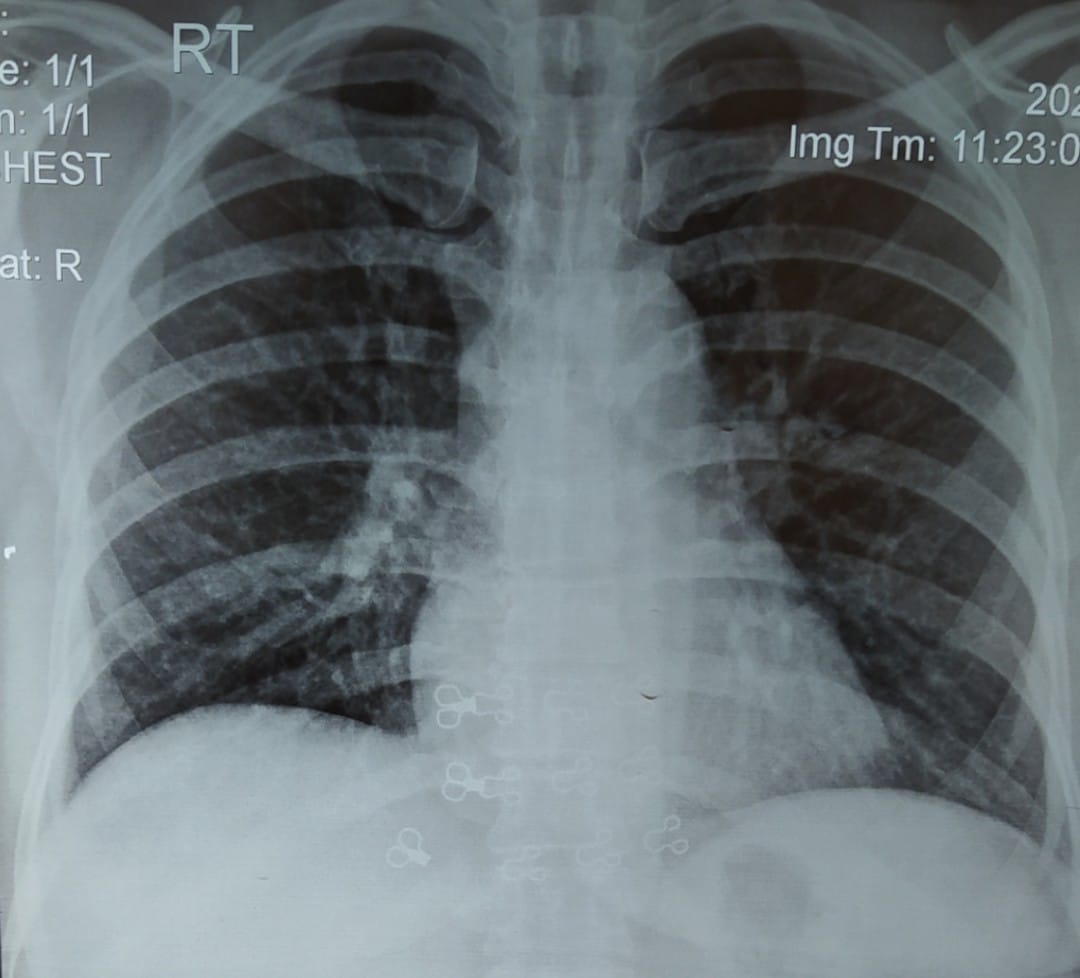
Perform a chest X ray and look for lung fields and cardiac size.

Also check Urea and electrolytes, creatinine levels and any evidence of sepsis

Check Haemoglobin levels.

Check blood sugar levels

Send sepsis screen

Further management:

Patients with simple syncope may be discharged

Those with underlying causes / Seizures or cardiac causes shpuld be admitted and investigated further.

Wednesday, 13 October 2021

TCA POISONING / TRICYCLIC ANTIDEPRESSANT POISONING :

 TCA POISONING / TRICYCLIC ANTIDEPRESSANT POISONING :

This is usually caused by Tri cyclic Antidepressants poisoning like

Amitryptiline

Imipramine

Dothiepin

But it may also occur due to other medications such as Atropine and Procyclidine.

Atropine is also present in Atropa Belladona ( deadly Nightshade )

Clinical Features :

Common Features include :

Tachycardia

Hot Dry skin

Dry mouth

Drowsiness leading to Coma

Dilated pupils

Ataxia

Urinary retention

Jerky limb movements.

Unconscious patients will have divergent squint ,

Increased muscle tone

Increased Reflexes,

Myoclonus and

Extensor plantar responses.

In deep coma, there will be muscle flaccidity with no detectable reflexes and respiratory depression requiring IPPV.

Convulsions occur in 10 percent of unconscious patients and may precipitate cardiac arrest.

Patients recovering from coma often suffer  delirium and hallucinations and have jerky limb movements and severe dysarthria.

ECG Findings :

Sinus tachycardia is usual.

But as the severity of poisoning increases, the PR interval and the QRS complex also increases.

Theese changes may help  confirm the clinical diagnosis of tricyclic poisoning in an unconscious patient.

The p wave maybe superimposed on the preceding  T wave giving the impression of VT  when the rhythm is actually sinus tachycardia with prolonged conduction.

In very severe poisoning ventricular arrhythmias and bradycardia can occur especially in patients who are hypoxic.

Death may result from cardiorespiratory depression and acidosis.

Management :

Clear Airway

Maintain ventilation.

Give supportive treatment and provide nursing care.

Observe continuously as the patient has the potential for rapid deterioration.

Monitor ECG during gastric lavage or unconscious patients or post ictal patients.

Perform gastric lavage if less than 1 hour since overdose or longer and if patient is in coma.

Activated charcoal should be given.

Single brief fits do not warrant any treatment but iv diazepam should be given in case of prolonged fits.

Most cardiac arrhythmias occur in the patients of TCA overdose while they are unconscious within a fe hours of overdose..

These can usually be treated in most cases by correction of Hypoxia and Acidosis.

8.4 % of sodium bicarbonate ( 50 – 100 ml in adult ) ( 1ml/kg in child ) may produce a dramatic improvement in cardiac rhythm and output.

This is achieved by altering protein binding and reducing active free Tricyclic drug.

Avoid anti arrhythmic drug

Donot use physostigmine salicylate  or flumazenil which ca precipitate fits.

Unconscious patients usually improve over 12 hours and rgain consciousness with in 36 hours

Delirium and hallucinations may persist over 2 -3 days

And may require sedation in large doses with diazepam ( 20 – 30 mg PO every 2-3 hours )


Wednesday, 6 October 2021

Aspirin / Salicylate Poisoning

 Aspirin / Salicylate Poisoning

One standard Aspirin tablet contains 300 mg of Acetyl salicylic acid

Ingestion of 150 mg/kg body weight produces mild toxicity.

Ingestion of 500 mg/kg body weight produces severe toxicity and fatal poisoning.

Ingestion of salicylate ointment from the skin can also produce toxicity.

Clinical features of Salicylate poisoning:

Tinnitus

Hyperventilation

Vomiting

Deafness

Sweating

Vasodilatation

Dehydration

Hypokalemia

In severe poisoning

Coma

Convulsions

Confusion

Children usually develop :

Hyperpyrexia and

Hypoglycemia

Rarer features include :

Non cardiogenic pulmonary oedema

Renal failure and 

Cerebral oedema

Metabolic and Acid Base features :

These include mixed Metabolic acidosis and Respiratory Alkalosis

But respiratory features predominate.

In a few childreand and adults , acidosis predominates and is often associated with  confusion or coma.

Management

Gastric lavage if the adult had ingested more than 4.5 Gm of Acetyl salisylic acid ( 15 tablets ) in the last 1 hour.

If the adult has ingested more than 4.5 Gm of Acetyl salicylic acid, 50 Grams of harcoal should be put in the stomach via the NG tube to reduce the absorption of Acteyl salicylic acid and to increase its elimination.

In children , if they have ingested more than 2 Grams , 25 Grams of charcoal should be put into the stomach.

Plasma salicylate concentration should be measured and repeated aftera few hours again as well.

This is because salicylate levels may increase due to continuing absorption.

In case of signs of severe poisoning or CNS Features , Plasma glucose levels, Urea and Electrolytes and Arterial Blood Gas levels should also be checked.

Mild poisoning :

Childern with plasma salicylate of less than 350 mg /l ( 2.5 mmol/l ) and adults with plasma salicylate levels of less than 450 mg /l ( 3.3mmol/l )  only need increase oral fluids to treat raised salicylate levels.

Moderate poisoning:

Children with salicylate levels of more than 350 mg/l and adults with salicylate levels of more than 450 mg/l will need iv fluids for correction of dehydration and elimination of salicylate.

Measure plasma salicylate levels and measure after a few hours again if symptoms recur as salicylate absorption may be ongoing.

Sodium bicarbonate 1.26 % ( 500 ml /hour , 3 hourly ) can correct metabolic acidosis and increase the elimination of salicylate as well as it alkanizes the urine and is superior to massive diuresis in elimination of salicylates.

Urinary pH should be more than 7.5 ( ideally 8.0 – 8.5 )

Repeated doses of Charcoal should be given.

Check Urea & Electrolytes, ABGs and serum potassium levels.

Repeat salicylate levels as needed.

Get urgent specialist  advice.

Consider urgent referral for Haemodialysis.

Salicylate Levels of more than 700 mg/L or 5.1 mmol/litre , CNS features are suggestive of severe toxicity and warrant seeking of urgent specialist advice and Haemodialysis.

Correct acidosis and give repeated charcoal by N/G tube.

IPPV may help in extreme hyperventilation with paralysis and and in life threatening poisoning with Coma

Haemodialysis removes salicylates and corrects electrolyte Imbalances.

Give additional glucose since brain glucose maybe low despite normal blood glucose concentrations.

In life threatening  poisoning with coma and extreme hyperventilation paralysis , IPPV may help while haemodialysis removes salicylates and corrects the electrolyte disturbances.


Friday, 3 September 2021

A 45 year old female presents with increasing shortness of breath and bilateral pitting oedema of both legs.

 A 45 year old female presents with increasing shortness of breath and bilateral pitting oedema of both legs. Her oxygen saturation is 86 percent. She is afebrile and her inflammatory markers are normal.

BP :145/65 mmHg


Her Chest X-ray is given below:

1. What are the chest X-ray findings?

2. What is the most likely Diagnosis?

3. What further investigation Should be performed?

4. How will you manage this condition?


Answers given in comments section.

Copyright reserved with the author.


1. What are the chest X-ray findings?
'Cardiomegaly+ peri hilar congestion + Bilateral pleural effusions.

2. Most likely Diagnosis: 
Advanced Congestive Cardiac Failure

3. Further investigations :

Echocardiogram

BNP levels


4. Management:

Intravenous diuresis with furosemide 40 mg 8 am, 20 mg 2 pm

High flow hoxygen

Bisoprolol

Loprin

Clopidrogel


https://www.printful.com/a/3070223:bc183f89d5ad9788fb4ac6ca2d069764

https://www.medstudentnotes.com/?ref=SAADSHAH

https://rebrand.ly/TeamToko?p=1564-31

https://urownceo.net/1564-25.php

Saturday, 7 August 2021

A 45 year old man presents to your clinic with complaints of increasing shortness of breath over last 6 months.

 A 45 year old man presents to your clinic with complaints of increasing shortness of breath over last 6 months.

He also gets occasional episodes of dry cough when walking or going up stairs.

On examination :

He is afebrile / normal Temperature and oxygen saturation is 89 Percent on air .

You organise a chest X ray which is given below:


1. What are findings on chest X ray?

2. What is the most likely Diagnosis?

3. What are the causes of this condition?

4. What further investigations would you like to perform?

5. How would you manage this condition?


Copyright reserved with author.

Answers given in comments section. 


1. What are findings on chest X ray? 

Increased interstitial markings” on chest x-ray because of the inflammation, swelling or scarring of the interstitium makes the tissue denser so that it is now visible as white “interstitial markings” on the x ray or scan.

2. What is the most likely Diagnosis? 

Interstitial Lung Disease (ILD) Interstitial Lung Disease (ILD) refers to a group of problems in the lung that affects the “interstitium”. 

The interstitium refers to the tissue area in and around the wall of the airsacs (alveoli) of the lung area where oxygen moves from the alveoli into the the capillary network (small blood vessels) that covers the lung like a thin sheet of blood. 

Once the oxygen crosses the intersitial space it enters the bloodstream and is delivered to the vital organs of your body. ILDs cause this interstitial space to become inflammed or scarred making it more difficult for oxygen.


3. What are the causes of this condition ? Causes of ILD

There are many causes of ILDs and they can be categorized as pictured above into three broad, main groups: 

1) Exposure-Related, 

2) Autoimmune-Related and 

3) Idiopathic, or with no known cause. Exposure-Related (Hypersensitivity Pneumonitis or 'HP'):

Inhalation-related:

There are more than 300 causes of inhalational exposure-related ILDs.

These exposures can be experienced in the workplace, at home, while doing hobbies, or during travel.

Some of the more common exposures are mold, birds in or around the home, bedroom, or backyard, farming/agricultural work, indoor hot tubs, standing water, down feathers, and cleaning chemicals.

Historically, coal miners (pneumoconiosis) and exposure to asbestos (asbestosis) were significant problems, but these exposures have lessened due to the use of protective respiratory equipment.

Medication-related: Some of the medications that can cause an inflammatory reaction in the lung include amiodarone, methotrexate, some chemotherapy agents used to treat cancers, and nitrofurantoin (sulfa drugs). 


Autoimmune-Related ILDs (Nonspecific Interstitial Pneumonitis – NSIP): Autoimmune–related ILDs tend to occur more often in women and in persons under the age of 65. Idiopathic ILDs (no known cause):

Idiopathic Pulmonary Fibrosis (IPF) is the most common ILD that has no known cause.


4. What further investigations would you like to perform? Radiology evaluations:

A Computed Tomography (CT) scan of the chest is the most sensitive diagnostic tool for ILD. The best assessment is with a high resolution CT scan (HRCT) which provides thin slices (1-2 mm thick) of the lungs in order to closely evaluate the lung tissue changes. Chest X-Rays may show ILD but are not diagnostic. Breathing tests:

Pulmonary Function Studies (PFTs) are used to evaluate the severity of the lung problem. PFTs evaluate the ability of the lung to:

move air in and out of the lungs (spirometry- Forced Vital Capacity (FVC), Forced Expiratory Volume in 1 second (FEV1). Lung Tissue Sampling: If the decision is made that a lung tissue sample (lung biopsy) is needed in order to make a diagnosis, there are a few different options:

Transbronchial Biopsy (TBBx) with Bronchoalveolar Lavage (BAL) can be done to obtain a very small piece of lung tissue and also a sample of a ‘washing’ of sterile water in and out of the lungs. It is performed as an outpatient procedure. 

Blood Tests: 

There are different types of blood tests that may be ordered to assist in making an accurate diagnosis Serology: These are blood tests that look for antibodies (proteins made by your immune system) in blood to determine if patient have a type of autoimmune disease (lupus, slceroderma, Sjögren's sydrome, polymyositis/ dermatomyositis, mixed connective tissue disease).

Examples of these blood tests include:

Rheumatoid Factor: rheumatoid arthritis

ESR: estimated sedimentation rate

CRP: C-reactive protein reflecting a response to inflammation

ANA: Antinuclear Antibodies

anti-dsDNA: anti-double stranded DNA

anti-SSA (Ro) and anti-SSB (La): anti Smith antigens

Anti-JO-1

ANCA: antineutrophil cytoplasmic antibodies

anti-Scl 70: scleroderma


5. How would you manage this condition? 

Pharmacological Treatment to suppress further inflammation includes medications such as : Corticosteroids /Prednisolone , Azathioprine, Cyclophosphamide, Hydroxychloroquine and Methotrexate. 

Anti-Fibrotic (anti-scarring) Agents include medications like Nintedanib (Ofev®) and Pirfenidone (Esbriet®, Pirfenex®, Pirespa®. 

Non-pharmacological options include : Pulmonary Rehabilitation , oxygen and Palliative Treatment.

 Pulmonary Rehabilitation (PR) is a structured exercise and education program for people with chronic lung diseases, including ILD, with the goal of maximizing a patient’s ability to maintain activity, decrease breathlessness and fatigue, and improve quality of life. 

Typically, pulmonary rehabilitation will include exercise training (aerobic, strengthening, flexibility); breathing exercises; anxiety, stress, and emotional management strategies; nutritional counseling; disease education; medication education, and other components.

https://drive.google.com/file/d/1M2Oi6_m0GhXR2KN7E43HcdwYCU59cIFQ/view?usp=sharing


https://drive.google.com/file/d/1M2Oi6_m0GhXR2KN7E43HcdwYCU59cIFQ/view? usp=sharing


Thursday, 5 August 2021

The CHA2DS2-VASc score

 Although simple, the CHADS2 score does not include many common stroke risk factors, and its limitations have been highlighted by its non-inclusion of common stroke risk factors. Even patients classified as low risk by CHADS2 in its original validation study have a stroke rate of 1.9% per year, which is close to the criterion of a cardiovascular event rate of 20% over 10 years for primary prevention strategies.

Consequently, CHADS2 was expanded to include three additional independent risk factors: vascular disease (coronary artery disease, peripheral artery disease, aortic atherosclerosis), age 65-74 years, and female sex. 

This new, more inclusive scoring system is the CHA2DS2-VASc score. 


The CHA2DS2-VASc score better discriminated stroke risk in nonvalvular AF subjects with a baseline CHADS2 score of 0 to 1


Table 3. CHA2DS2-VASc Score and Risk Criteria 

Score

CHA2DS2-VASc Risk Criteria

1 point

Congestive heart failure

1 point

Hypertension

2 points

Age ≥75 years

1 point

Diabetes mellitus

2 points

Stroke/Transient Ischemic Attack/Thromboembolic event

1 point

Vascular disease (prior MI, PAD, or aortic plaque)

1 point

Age 65 to 74 years

1 point

Sex category (ie, female sex)